
22 April 2026
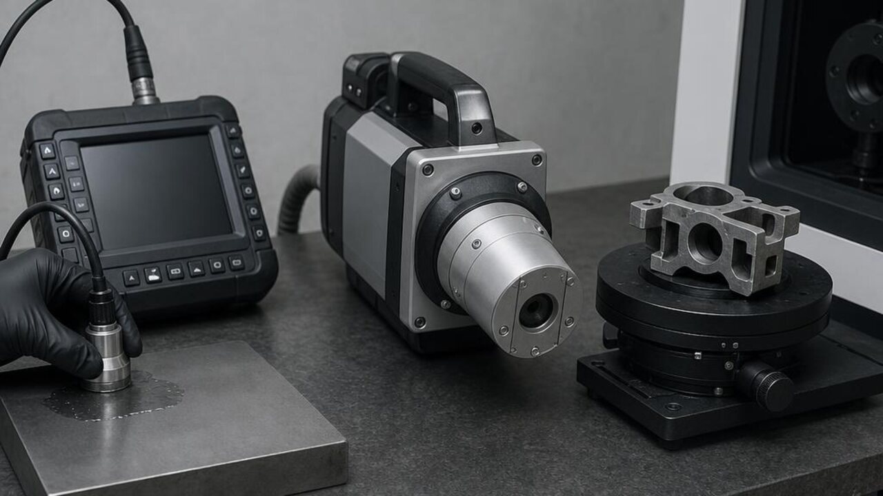
Three ways of seeing inside a part
X-ray radiography projects a two-dimensional image of a component's internal structure. Computed tomography (CT) rotates part or source to reconstruct a full three-dimensional volume. Ultrasonic testing sends sound waves through the material and reads their reflections. All three see beneath the surface; they differ in what they resolve, what they cost to operate and where they can physically go.
Where each method fits
Radiography excels at welds, castings and assemblies where a 2D projection reveals the defect classes of interest. CT is the reference method when internal geometry matters in three dimensions: complex castings, additively manufactured parts, electronics assemblies. Ultrasonics dominate field and in-service inspection: thickness measurement, corrosion mapping and flaw detection on structures too large or too installed to bring to an imaging bay.
The practical trade-offs
Radiographic methods need radiation-safety infrastructure and trained, certified operators; CT adds reconstruction time and fixturing discipline. Ultrasonics are portable and safe but demand skilled interpretation and access to the right surfaces. The choice is rarely either/or: production lines commonly pair radiography or CT for acceptance with ultrasonics for in-service follow-up.
Standards decide, not preference
In defence and aerospace work, the inspection standard usually settles the method question: acceptance criteria are written against specific techniques, coverage and sensitivity. Buyers should select equipment against the standards their programmes must satisfy, and confirm the supplier can demonstrate compliance on representative parts before commitment.
The bottom line for programme owners
For defence and critical-infrastructure buyers, the method debate is a distraction if it is settled by preference rather than by standard. The real advantage comes from letting the applicable acceptance criteria drive the choice, then building a toolkit that combines volumetric and in-service methods so nothing that could threaten the mission goes unseen. Radiography, CT and ultrasonics each protect a different part of the picture; mature programmes specify them in concert. Ask the supplier to prove compliance on representative parts before commitment, and secure the training, calibration and service that keep that compliance intact for years. Where in-region capability matters, localisation arrangements are scoped per programme, subject to export controls and end-use approvals, under one accountable channel rather than a scatter of point purchases.
